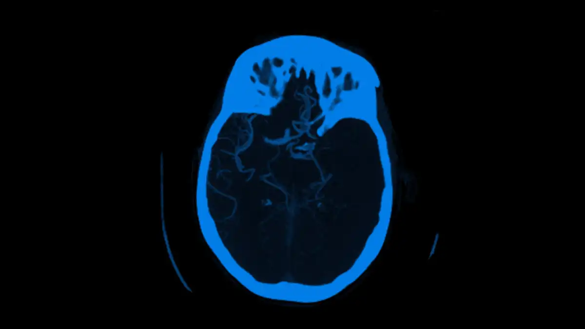
Viz™ LVO
AI software detection of large vessel occlusion stroke on...
Background Artificial intelligence (AI) software is increasingly applied in stroke diagnostics. However, the actual performance of AI tools for identifying...
Jan 27, 2022

Machine learning algorithms have shown groundbreaking results in neuroimaging. Herein, the authors evaluate the performance of a newly developed convolutional neural network (CNN) to detect and quantify the thickness, volume, and midline shift (MLS) of subdural hematoma (SDH) from noncontrast head CT (NCHCT).
NCHCT studies performed for the evaluation of head trauma in consecutive patients between July 2018 and April 2021 at a single institution were retrospectively identified. Ground truth determination of SDH, thickness, and MLS was established by the neuroradiology report. The primary outcome was performance of the CNN in detecting SDH in an external validation set, as measured using area under the receiver operating characteristic curve analysis. Secondary outcomes included accuracy for thickness, volume, and MLS.
Among 263 cases with valid NCHCT according to the study criteria, 135 patients (51%) were male, the mean (± standard deviation) age was 61 ± 23 years, and 70 patients were diagnosed with SDH on neuroradiologist evaluation. The median SDH thickness was 11 mm (IQR 6 mm), and 16 patients had a median MLS of 5 mm (IQR 2.25 mm). In the independent data set, the CNN performed well, with sensitivity of 91.4% (95% CI 82.3%–96.8%), specificity of 96.4% (95% CI 92.7%–98.5%), and accuracy of 95.1% (95% CI 91.7%–97.3%); sensitivity for the subgroup with an SDH thickness above 10 mm was 100%. The maximum thickness mean absolute error was 2.75 mm (95% CI 2.14–3.37 mm), whereas the MLS mean absolute error was 0.93 mm (95% CI 0.55–1.31 mm). The Pearson correlation coefficient computed to determine agreement between automated and manual segmentation measurements was 0.97 (95% CI 0.96–0.98).
The described Viz.ai SDH CNN performed exceptionally well at identifying and quantifying key features of SDHs in an independent validation imaging data set.
Researcher:
Marco Colasurdo , MD
Publication:
Journal of NeuroInterventional SurgeryDate Published: September 30, 2022
Sep 30, 2022