Viz™ AORTIC
A Review of Mature Machine Learning and Artificial Intelligence...
Background After years of both increasing enthusiasm and skepticism by surgeons and patients, the first artificial intelligence (AI) and machine...
May 05, 2023
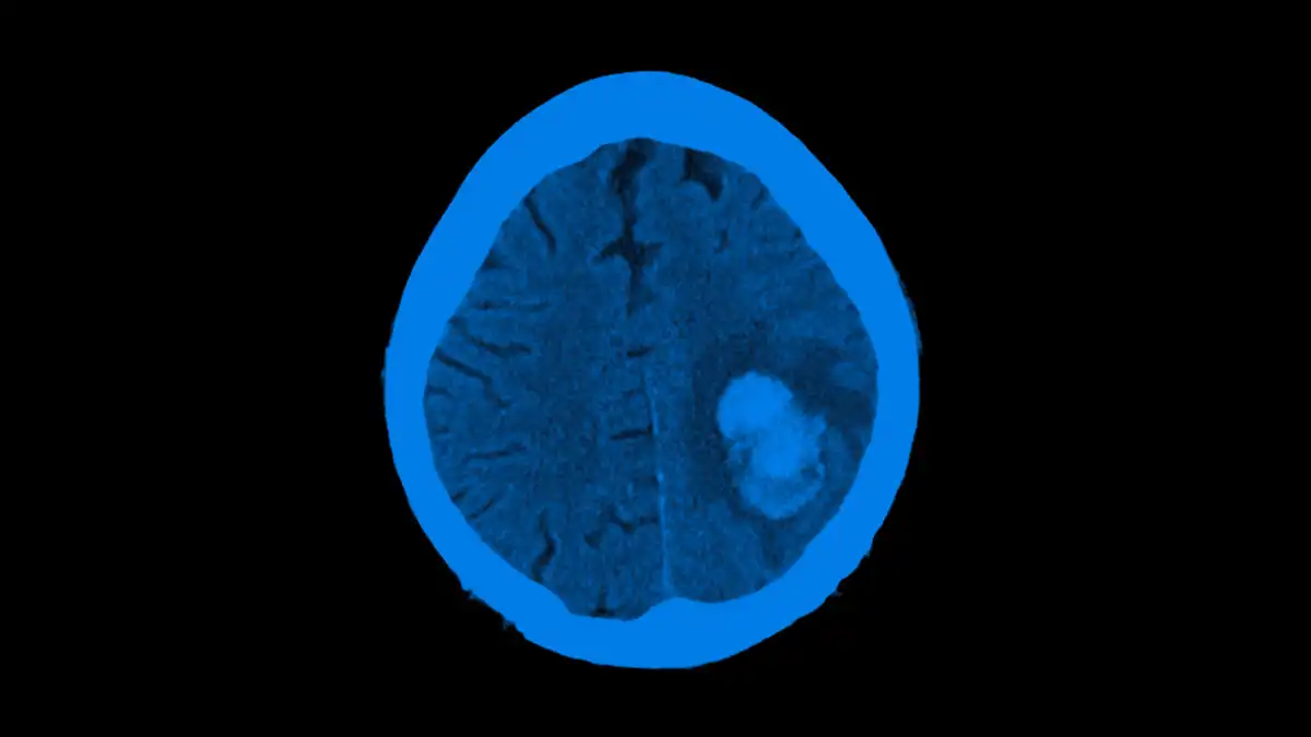
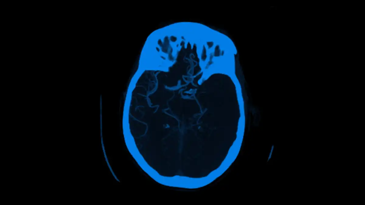
The diagnosis of stroke is based on the clinical examination and also on different imaging technics. The differentiation between haemorrhagic and ischemic stroke and the detection of large vessel occlusions (LVO) represent key steps in determining the optimal therapy regimen for the individual patient. Time is essential: the faster the diagnosis is made, and the appropriate therapy is initiated, the better the outcome of patients [1]. Many tools for medical image analysis have been developed to reduce the time needed to detect abnormalities and to provide more accurate results. Particularly, tools based on machine learning techniques have led to significant improvements in medical imaging interpretation in the last decade.
In the 1980s, a concept called computer-aided diagnosis (CAD) was introduced. Its primary intent was to provide radiologists with a second opinion while reading their cases [2]. CAD has seen remarkable developments, and applications for all modalities of medical imaging have been presented. Although CAD systems are widely available, their implementation in clinical routine varies with the clinical scenario in which they are applied. In the first years following the emergent of CAD concept, the majority of the developed algorithms focused on the early detection of breast cancers on mammograms and the detection of lung cancer on chest radiographs or computed tomography. Currently, CAD systems are well established for providing an aid diagnosis in stroke and many other medical fields (Additional file 1).
Automated image analysis of ischemic stroke with the support of machine learning or artificial intelligence related algorithms is a constantly growing market. Commercially and non-commercially available CAD products so far focus on the analysis of NCCT, CTA and perfusion imaging, based on CT or MR imaging. They aim to identify and quantify the ischemic core, the ischemic penumbra, the status of collateral flow and the site of arterial occlusion in an automatic fashion.
CAD algorithms are not intended as standalone diagnostic tools, however, they assist physicians to get more accurate and standardised interpretations of stroke related findings, which may improve the stroke management and patients’ selection for appropriate (usually time critical) treatments.
Future clinical studies are necessary for proper validation, evaluation and comparison of the different available software solutions in order to broaden and generalise treatment selection criteria for patients with acute ischemic stroke. Furthermore, future studies may focus on the integration of CAD algorithms within the workflow of stroke referral networks.
Researcher:
Dr. Yahia Mokli
Publication:
Biomed CentralDate Published: August 15, 2019
Aug 15, 2019
Viz™ AORTIC
Background After years of both increasing enthusiasm and skepticism by surgeons and patients, the first artificial intelligence (AI) and machine...
May 05, 2023
Viz™ LVO
Background Artificial intelligence (AI) software is increasingly applied in stroke diagnostics. However, the actual performance of AI tools for identifying...
Jan 27, 2022
Viz™ LVO
Background Prompt and accurate identification of Large Vessel Occlusion (LVO) for Acute Ischemic Stroke (AIS) by CTA is essential for...
Jan 30, 2019